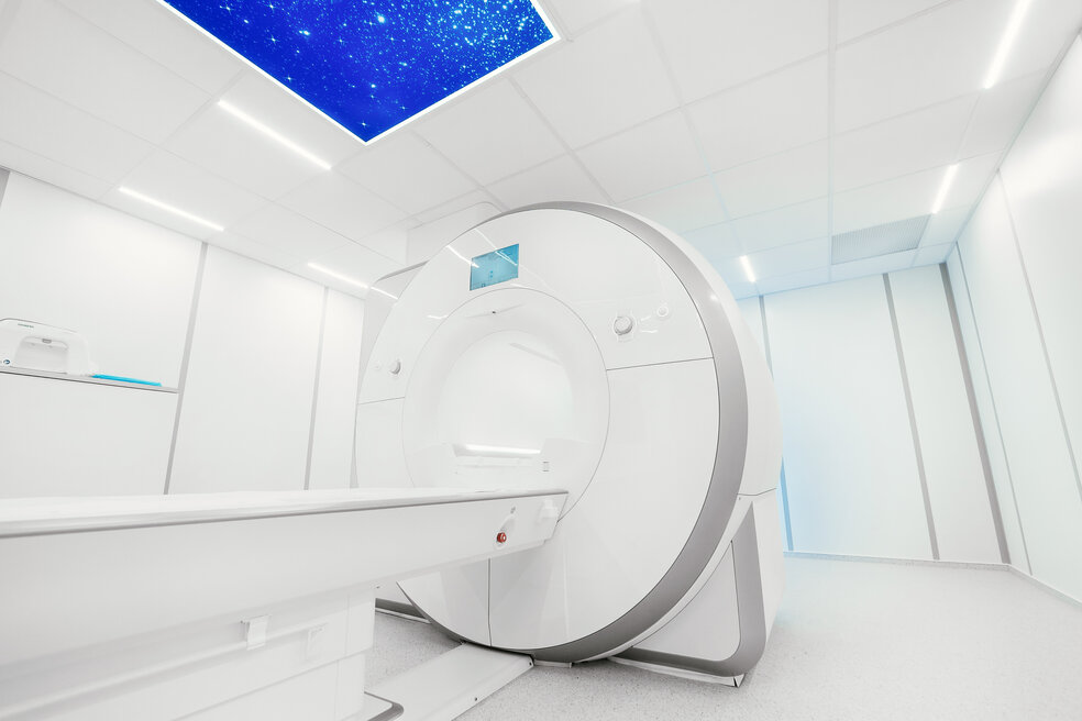
Nuclear medicine imaging is gaining more and more attention in the field of medical imaging. It has proven useful in many instances. It is used in adults to detect and treat anomalies in the heart, lungs, brain, bones and other systems.
Nuclear medicine imaging uses radiopharmaceuticals or radiotracers which are injected, swallowed, or inhaled depending on the test. These contain small amounts of radioactive material that can be detected from a Positron Emission Tomography (PET) scan. These radiotracers commonly use a molecule similar to glucose called F-18 fluorodeoxyglucose or FDG. These molecules get collected in more metabolically active regions like cancer cells due to their similarity to glucose. With accumulation, they emit gamma rays, which can be detected by a gamma camera. This accumulation can take mere seconds or even days. Depending on this, the imaging may be done accordingly. The time-consuming nature is often considered as the main limitation of the Nuclear medicine imaging procedure.
The gamma camera contains radiation detectors. These cameras are attached to the gantry to detect gamma rays emitted from the radiotracer in the body. Then, the camera sends its data to the computer to generate an image. This process is carried out using a PET scanner or a handheld probe. While the PET scanner helps to handle full body scans, the probe is useful in measuring the radiation emitted from a smaller, specific area. Nuclear medicine imaging superimposed with CT is brought to us as PET/CT. PET/MRI, which brings together PET and MRI is also an emerging technology in this field.
When it comes to radioactive materials, the associated risk is a mandatory topic of discussion. But in this case, the risk is very low compared to the benefits as only a minimal dose is administered. The radioactive material left in the body will lose its radioactivity with time following the process of radioactive decay. It is important to drink lots of water following the test to flush out amounts of radiotracer with urine.
Allergic reactions to the radiotracer, although rare, have only mild effects. But it is essential to inform about any pregnancies, confirmed or suspected, breastfeeding and any other conditions including allergies and other medications.
Researchers continue to make advances in the field of nuclear imaging in terms of diagnosis, treatment, and even monitoring of the effectiveness of treatment. The benefits of nuclear medicine, more or less seem to outweigh the risks associated.
The benefits and the quality of medical imaging services mostly depend on the medical imaging equipment used in the procedure. If you are looking to purchase high-quality medical imaging equipment (GE X Ray Equipment For Sale) for sale, you have come to the right place. With us here at Amber Diagnostics you are guaranteed the best medical imaging equipment, used and refurbished at the best prices available in town. Call us up to get in touch with our agents to find out more about our equipment. If you need help selecting the right medical imaging equipment for your facility, we can even help you out with that. With decades of experience in the market, you can trust Amber Diagnostics to be your medical imaging equipment provider. Call our hotline now to find out more!
![Nuclear medicine imaging is gaining more and more attention in the field of medical imaging. It has proven useful in many instances. It is used in adults to detect and treat anomalies in the heart, lungs, brain, bones and other systems. Nuclear medicine imaging uses radiopharmaceuticals or radiotracers which are injected, swallowed, or inhaled depending […]](https://www.sinky.net/wp-content/uploads/2018/01/Untitled-1-1.png)


