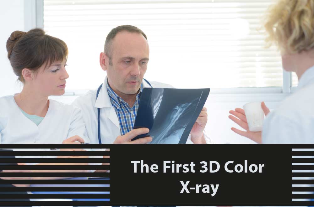
What if instead of black and white images, a medical imaging practitioner was able to acquire colored images on the tissues being scanned by an x-ray? This color imaging technique would also be able to produce more vivid and accurate pictures within the body. This technique would then be able to provide doctors and patients with a more accurate diagnosis of the internal structure of the body.
Fortunately, this has become a reality in some countries. This was one of the most significant breakthroughs in the technological and scientific industry. A company in New Zealand known as Mars Bioimaging has developed a new type of medical imaging scanner that allows x-rays to see inside the human body as a typical x-ray. However, the difference is that these developers have borrowed a technology developed for the Large Hadron Collider at CERN which provided improved results. The use of this scanner also helps differentiate bone, muscles, fat, liquids, and other materials inside the body. Additional software used for the device helps to produce stunning full-color images that allow a 3D view of the inside of the body.
So this means, with the current developments, while a doctor is examining images of a patients arm or leg for a nasty fall, the practitioner would also be able to observe any other potential medical conditions that might not be visible in typical x-ray systems. This new technology can be used in countless medical fields from dentistry to brain surgery.
While it might take years before the new system receives the clearances and approval to be used in hospitals, this breakthrough will certainly change the world. Until then, developers are also seeking to make improvements in the current standard of x-ray devices.
Some of the best used and refurbished x ray equipment in the world can be purchased through Amber USA. They guarantee a variety of options to cater to your medical facility’s needs while guaranteeing optimal outcomes from all the equipment. Contact us now for inquiries!
![What if instead of black and white images, a medical imaging practitioner was able to acquire colored images on the tissues being scanned by an x-ray? This color imaging technique would also be able to produce more vivid and accurate pictures within the body. This technique would then be able to provide doctors and patients […]](https://www.sinky.net/wp-content/uploads/2018/01/Untitled-1-1.png)


